
Other SV Defects

While most families in the Sisters by Heart community are impacted by the single ventricle defect Hypoplastic Left Heart Syndrome, there are several other congenital heart defects resulting in single ventricle diseases. These include:
Tricuspid Atresia (TA)
Double Inlet Left Ventricle (DILV)
Double Outlet Right Ventricle (DORV)
Atrioventricular Canal Defect (AV)
Pulmonary Atresia with Intact Ventricular Septum (PA-IVS)
These defects often result in a single ventricle heart anatomy via the Fontan surgery.
For more information, please visit:
https://www.cincinnatichildrens.org/health/s/sv
https://www.chop.edu/conditions-diseases/single-ventricle-heart-defects

Tricuspid Atresia
In tricuspid atresia, blood can't flow from the right atrium to the right ventricle because the valve between them is missing. This condition often includes a smaller than normal right ventricle, as well as an atrial septal defect and, in some cases, a ventricular septal defect.
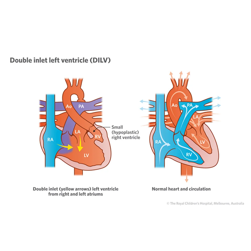
Double Inlet Left Ventricle
Normally, the tricuspid valve leads into the right pumping chamber (ventricle), and the mitral valve leads into the left ventricle. These valves act as 1-way doors, allowing blood to flow from the atria to the ventricles without letting blood back into the atria. A wall called the septum separates the ventricles. In babies with this defect, both the tricuspid valve and the mitral valve let blood flow into the left ventricle. Only the left ventricle works. Because the right ventricle is not being used, it is small and not well developed. This is also called a single-ventricle heart defect because children with this defect only have 1 ventricle in their heart that works. Single-ventricle defects are some of the most complex heart-related birth defects.
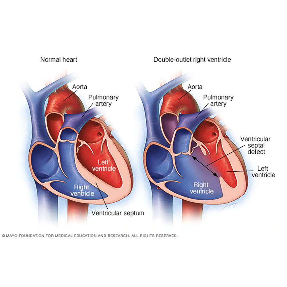
Double Outlet Right Ventricle
In double-outlet right ventricle, the aorta and pulmonary artery connect partially or completely to the right ventricle. A hole also exists between the two ventricles (ventricular septal defect). In a normal heart, as shown on the left, the pulmonary artery connects to the right ventricle and the aorta connects to the left ventricle.
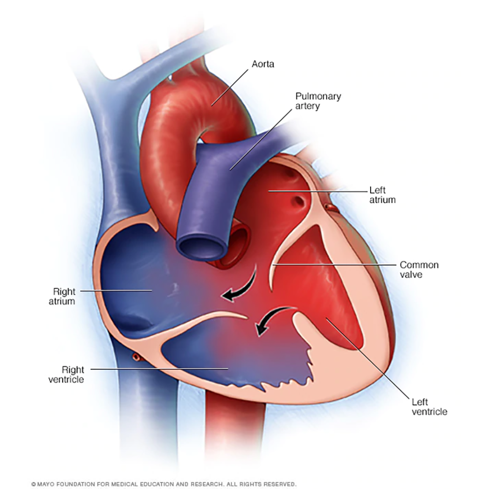
Atrioventricular Canal Defect
Atrioventricular canal defect includes a hole in the wall between the heart's chambers and flaws in the heart's valves. This defect is classified by whether it's partial, involving only the upper two chambers, or complete, in which blood can travel freely among all four chambers.
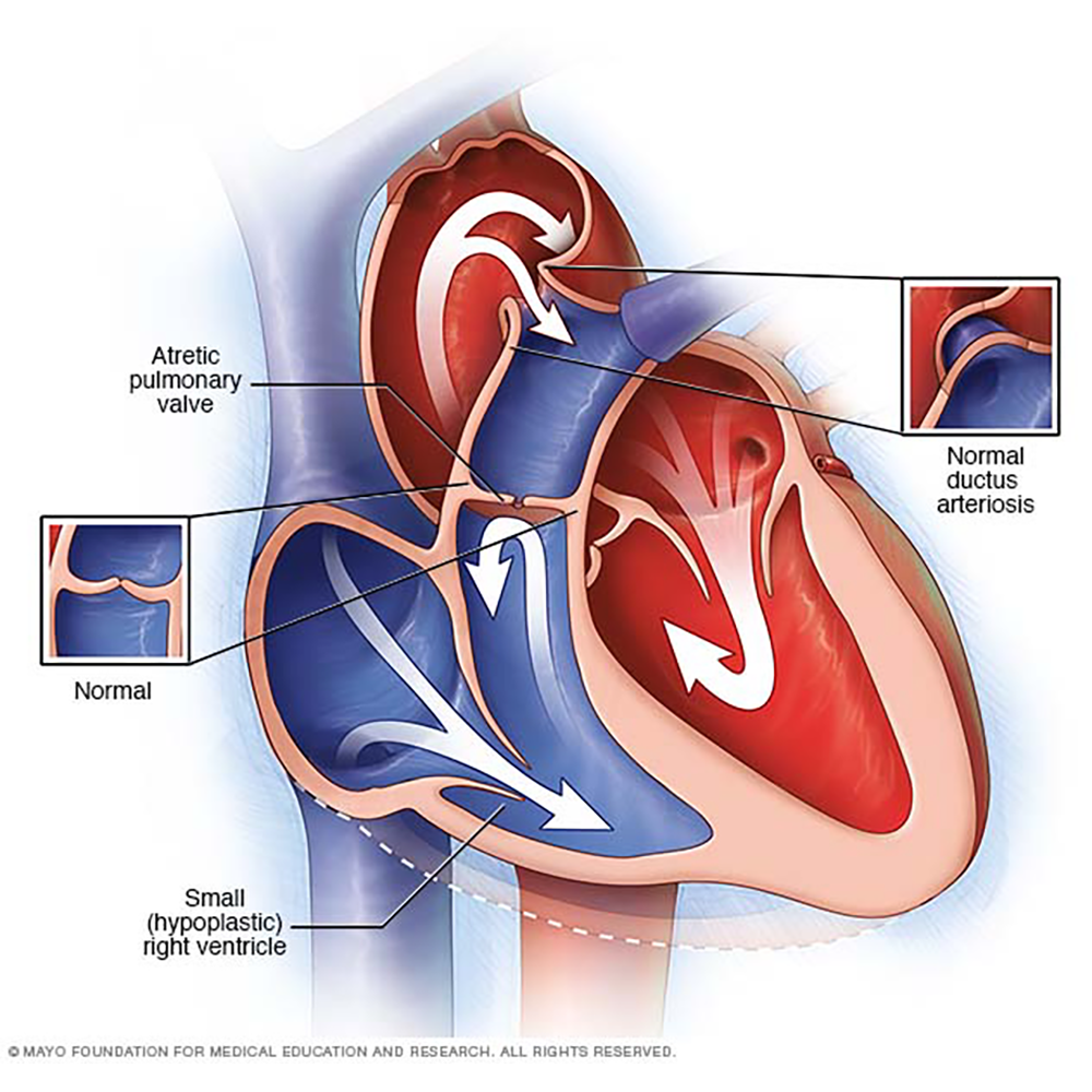
Pulmonary Atresia with Intact Ventricular Septum
In pulmonary atresia, the valve that lets blood flow from the heart to the lungs isn't formed properly. Instead, some blood may reach the lungs via a temporary connection (ductus arteriosus) between a baby's aorta and the pulmonary artery. Some babies born with pulmonary atresia may have a small (hypoplastic) right ventricle.